Abstract
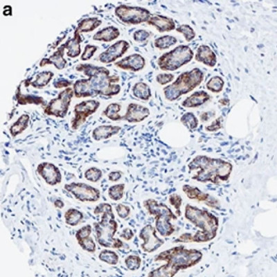
P-cadherin is a calcium-dependent cell-cell adhesion glycoprotein. P-cadherin expression is restricted to the myoepithelial cells in normal breast tissue, and aberrant staining has also been described in invasive tumors. Several small studies have reported P-cadherin as a marker of poor outcome in breast cancer patients but its prognostic significance in relation to other variables has not been established in a large series of breast cancers. A tissue microarray was constructed from 3992 cases of invasive breast carcinoma, and P-cadherin expression was evaluated using immunohistochemistry. Median follow-up was 12.5 years. The immunohistochemistry-based definitions of cancer subtypes were luminal (ER+ or PR+/HER2-), luminal/HER2+ (ER+ or PR+/HER2+), HER2+ (ER-/PR-/HER2+), and basal (ER-/PR-/HER2-/CK5/6+ or EGFR+). Clinical covariate and biomarker associations were assessed using contingency tables, and Pearson’s χ(2) or Fisher’s exact test. Survival associations were assessed using Kaplan-Meier plots, logrank and Breslow tests, and Cox proportional hazards regression analysis. P-cadherin was expressed in 34.8% (1290/3710, 50% cut point) of cases. P-cadherin staining was strongly associated with HER2+ and basal carcinoma subtypes (P<0.0005). P-cadherin-positive patients showed significantly poorer short-term (0-10 years) overall survival, disease-specific survival, distant relapse-free interval, and locoregional relapse-free interval in univariable models (P<0.05). In multivariable Cox models containing standard clinical covariates and cancer subtypes, P-cadherin did not show independent prognostic value. P-cadherin expression was positively associated with histological grade, chemotherapy, Ki-67, EGFR, CK5/6, p53, YB-1, and HER2 expression (P<0.002), and negatively associated with age at diagnosis, ER, PR, and Bcl-2 expression (P<0.0005). This study shows the value of P-cadherin as a marker of poor prognosis. The large sample size of this series clarifies contradictory findings of many smaller studies. P-cadherin positivity is associated with high-grade tumor subtypes and well-established markers of poor prognosis, and may represent a promising antibody therapeutic target.