Abstract
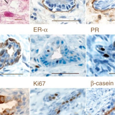
Previous studies have demonstrated that normal mouse mammary tissue contains a rare subset of mammary stem cells. We now describe a method for detecting an analogous subpopulation in normal human mammary tissue. Dissociated cells are suspended with fibroblasts in collagen gels, which are then implanted under the kidney capsule of hormone-treated immunodeficient mice. After 2-8 weeks, the gels contain bilayered mammary epithelial structures, including luminal and myoepithelial cells, their in vitro clonogenic progenitors and cells that produce similar structures in secondary transplants. The regenerated clonogenic progenitors provide an objective indicator of input mammary stem cell activity and allow the frequency and phenotype of these human mammary stem cells to be determined by limiting-dilution analysis. This new assay procedure sets the stage for investigations of mechanisms regulating normal human mammary stem cells (and possibly stem cells in other tissues) and their relationship to human cancer stem cell populations.
