Abstract
Mammographic density is the third largest risk factor for ductal carcinoma in-situ (DCIS) and invasive breast cancer. However, the question of whether risk-mediating precursor histological changes, such as columnar cell lesions (CCLs), can be found in dense but non-malignant breast tissues has not been systematically addressed. We hypothesized that CCLs may be related to breast composition, in particular breast density, in non-tumour containing breast tissue.
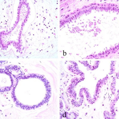
We examined randomly selected tissue samples obtained by bilateral subcutaneous mastectomy from a forensic autopsy series, where tissue composition was assessed, and in which there had been no selection of subjects or histological specimens for breast disease. We reviewed H&E slides for the presence of atypical and non-atypical CCLs and correlated with histological features measured using quantitative microscopy.
CCLs were seen in 40 out of 236 cases (17%). The presence of CCLs was found to be associated with several measures of breast tissue composition, including radiographic density: high Faxitron Wolfe Density (P = 0.037), high density estimated by percentage non-adipose tissue area (P = 0.037), high percentage collagen (P = 9.2E-05) and high percentage glandular area (P = 2E-05). DCIS was identified in two atypical CCL cases. The extent of CCL was not associated with any of the examined variables.
Our study is the first to report a possible association between CCLs and breast tissue composition, including mammographic density. Our data suggest that prospective elucidation of the strength and nature of the clinicopathological correlation may lead to an enhanced understanding of mammographic density and evidence based management strategies.
